Article Text

Abstract
Background Transbronchial cryoablation shows potential as a local therapy for inoperable peripheral lung cancer. However, its clinical application for peripheral pulmonary lesions has not been reported yet.
Methods An improved cryoprobe with an 8-mm-long, 1.9-mm-wide cryotip was used. Initially, the safety and effectiveness of this cryoprobe were assessed in an in vivo porcine model. Transbronchial cryoablation with 2 or 3 freeze-thaw cycles (10 min or 15 min in each freezing time) was performed in 18 pigs under CT monitoring. Radiological and pathological examinations were performed to evaluate the extent of cryoablation. Subsequently, nine patients with stage IA peripheral lung cancer or metastases underwent transbronchial cryoablation with this cryoprobe under the guidance of navigation bronchoscopy and cone-beam CT. Technical success, safety and outcomes were assessed.
Results 36 cryoablation procedures were performed successfully without any major complications in the porcine model. The extent of cryoablation increased with freezing time and the number of freeze-thaw cycles, which peaked at 24 hours and then gradually decreased. Pathological results showed a change from massive haemorrhage at 24 hours to fibrous hyperplasia with chronic inflammation after 4 weeks. In the clinical trial, 10 cryoablations were performed on 9 tumours with a technical success rate of 100%. One mild treatment-related complication occurred. Of the nine tumours, seven achieved complete ablation, while two exhibited incomplete ablation and subsequent local progression at 6 months.
Conclusion Our initial experience indicated that transbronchial cryoablation was a safe and feasible procedure for non-surgical peripheral stage IA lung cancer or pulmonary metastases.
Trial registration number ChiCTR2200061544.
- Bronchoscopy
- Lung Cancer
Data availability statement
Data are available upon reasonable request.
This is an open access article distributed in accordance with the Creative Commons Attribution Non Commercial (CC BY-NC 4.0) license, which permits others to distribute, remix, adapt, build upon this work non-commercially, and license their derivative works on different terms, provided the original work is properly cited, appropriate credit is given, any changes made indicated, and the use is non-commercial. See: http://creativecommons.org/licenses/by-nc/4.0/.
Statistics from Altmetric.com
WHAT IS ALREADY KNOWN ON THIS TOPIC
Local tumour cryoablation is recommended for non-surgical treatment modality of early-stage lung cancer. A transbronchial approach is a possible therapeutic alternative, with a reduced risk of pneumothorax. However, no bronchoscopy-guided cryoablation has been performed to date for peripheral pulmonary lesions in clinical practice.
WHAT THIS STUDY ADDS
Transbronchial cryoablation with a novel thin cryoprobe was successfully performed in an in vivo porcine model and tested in an exploratory clinical study of early-stage peripheral lung cancer and some pulmonary metastases.
HOW THIS STUDY MIGHT AFFECT RESEARCH, PRACTICE OR POLICY
This is the first report of transbronchial cryoablation for peripheral pulmonary lesions and supports further evaluation of this technique in adequately powered clinical trials.
Introduction
Surgical resection remains the recommended treatment for early-stage non-small cell lung cancer (NSCLC) and some pulmonary metastases.1–3 For patients ineligible for surgery, local tumour ablation, such as radiofrequency ablation (RFA), microwave ablation (MWA) and cryoablation, has been applied percutaneously in clinical practice. Cryoablation may possibly be better tolerated and potentially safer than heating modalities4 and may induce a greater postablation immune response.5 Attempts to treat malignant tumours with an application of low temperature date back to the mid-19th century when it was first applied by Dr James Arnott.6 However, it was not until a century later that reliable cryosurgical instruments were manufactured. Nowadays, stable cryoablation systems use high-pressure gas, such as nitrogen or argon, that can expand rapidly at room temperature to cool the cryoprobe to −130°C to −170°C by the Joule-Thompson effect.7
However, percutaneous thermal ablation procedures have a high risk of pneumothorax and haemorrhage.8 Transbronchial ablation may be a safer therapeutic alternative, with the potential to decrease these severe complications.9 To date, several transbronchial catheters for thermal ablation of peripheral lung tumours have been developed. Good safety and efficacy have been shown based on a limited number of participants.10–14 Therefore, further evaluation in larger cohorts is still warranted. Our team developed a 2.2-mm-wide flexible cryoprobe (Y2212, AccuTarget MediPharma (Shanghai) Co., Ltd) that fitted through bronchoscopy working channels and evaluated its feasibility and safety ex vivo and in vivo.15 16 However, to date, there has been no clinical application of bronchoscopy-guided cryoablation for peripheral pulmonary lesions.
Therefore, we developed a 1.9-mm-wide thin cryoprobe in this study. We aimed to demonstrate the safety and efficacy of transbronchial cryoablation using this novel improved cryoprobe in in vivo lung parenchyma of porcine models and further investigate the initial clinical utility of cone-beam CT (CBCT)-guided transbronchial cryoablation for peripheral pulmonary lesions.
Methods
Animals
18 normal pigs weighing 40–60 kg were used. The animals were housed in an experimental cage for an adaptation period of 1 week before the experiment. To rule out other lung diseases before the procedure, each pig was scanned using CT. All pigs were randomly divided into two groups based on the duration of freezing time (10 min and 15 min groups, n=9 for each group). Each pig underwent two and three freeze-thaw cycles in both lungs. Each group was randomly divided into subgroups according to sacrifice time (24-hour, 4-week and 3-month groups, n=3 for each group). They were anaesthetised with an intravenous injection of 4–6 mg/kg/hour of propofol (Hospira, Inc, Lake Forest, California, USA) after an overnight fast. Tracheal intubation was performed, and breathing was maintained under ventilator control (SAVINA, Dräger, Telford, Pennsylvania, USA) at a respiratory rate of 12–14 breaths per minute with a tidal volume of 8 mL/kg. After cryoablation, dexamethasone (5 mg) was administered once and cefuroxime for 3 days after treatment. The weight, health status, diet, behaviour, medication and other aspects were observed and recorded. All applicable international, national and/or institutional guidelines for the care and use of animals were followed.
Cryoablation system
All cryoablation procedures were carried out using the Cryotherapy System (AT-2020-G, AccuTarget MediPharma (Shanghai) Co., Ltd) (figure 1A), which used nitrogen (full pressure=2100 pounds per square inch) as the refrigerant. A novel improved flexible cryoprobe (Y1908, AccuTarget MediPharma (Shanghai) Co., Ltd) with an 8-mm-long, 1.9-mm-diameter cryotip and a 1.15-m-long, 1.9-mm-diameter flexible catheter was used (figure 1A,B). Online supplemental figure S1 shows the operating principle of this novel flexible cryoprobe. The catheter incorporates a vacuum insulation layer designed to prevent damage caused by cold and heat to non-targeted bronchial airways and the bronchoscope. Moreover, the novel cryoprobe could fit through the working channel of 2.0 mm or larger. Figure 1B shows the adaptability of this novel flexible cryoprobe to different models of bronchoscopes (BF-1T260, BF-260 and BF-P260F, Olympus, Tokyo, Japan; outer diameter 5.9 mm, 4.9 mm and 4.0 mm, respectively; working channel diameter 2.8 mm, 2.0 mm and 2.0 mm, respectively).
Supplemental material

(A) Cryotherapy system and novel thin flexible cryoprobe. (B) Adaptability of the novel flexible cryoprobe to different models of bronchoscopes (BF-1T260, BF-260 and BF-P260F). (C) Experimental overview. (D) Experimental grouping and procedures created with BioRender.com.
In vivo porcine models
All animals had a full-chest, thin-slice CT scan before cryoablation for preoperative planning. Bronchoscopy-guided cryoablation was carried out by an experienced bronchoscopy expert. Ablation positions were chosen at plausible tumour locations (bronchioles of grade 5 or above, approximately 2 mm in diameter). A flexible cryoprobe was guided to the target bronchus through the working channel of the bronchoscope (BF-260, Olympus), which was confirmed by a CT scan. All apparatuses were held on the CT bed to allow for real-time synchronous CT scanning (figure 1C). Each freeze-thaw cycle comprised 10 min or 15 min freezing followed by 5 min thawing. Two and three freeze-thaw cycles were performed in both lungs. The experimental grouping and procedures are shown in figure 1D.
Radiological examination
CT images were obtained before and after each freeze-thaw cycle to assess the ablation and complications. Additional CT examinations were performed at 24-hour intervals (36/36 sites), 48-hour intervals (24/36 sites), after 1 week (24/36 sites), after 4 weeks (24/36 ablations) and after 3 months (12/36 ablations). The target bronchus was reconstructed by manual rotation on imaging software (RadiAnt DICOM Viewer, V.2022.1.1, Medixant, Poland). The maximal long-axis diameter (DL) and perpendicular maximal short-axis diameter (DS) of the ablation zone immediately, 24 hours, 48 hours, 1 week, 4 weeks and 3 months after cryoablation were measured by three experienced doctors on the lung window setting. DL and DS at each time point were used to calculate ablation volume using the following equation17: Volume = π/6×DL×DS2.
Histological examination
The ablation zones were dissected along the major axis of the target bronchus. The DL and DS of the ablated zones were measured according to visual demarcation between ablated lesions and normal tissues. Ablation volumes on gross were also calculated using the equation above. After that, the lungs were stored in 10% formalin, embedded in paraffin, cut into 4-μm-thick slices and stained with H&E and Masson stain. Histological changes in the lung parenchyma, bronchi and vessels after cryoablation were observed in sections perpendicular to the long axis of the cryoprobe tract.
Cytokine assay
Peripheral blood was collected from pigs before cryoablation and immediately, 24 hours, 48 hours, 1 week, 4 weeks and 3 months after cryoablation. Plasma was isolated after centrifugation for the examination of C reactive protein (CRP) and proinflammatory cytokines including interleukin (IL)-1β, IL-2, IL-12, tumour necrosis factor (TNF)-α and interferon (IFN)-γ using Porcine CRP ELISA kit (NOVUS, #NBP2-67253) and Porcine Luminex Discovery Assay (R&D Systems, #LXSAPM-07).
Statistical analysis
The data are presented as mean±SD. Between-group differences were examined using one-way analysis of variance (ANOVA) with Bonferroni’s multiple comparison test for equal variance or Welch’s ANOVA with Dunnett’s T3 multiple comparison test for unequal variance. In all analyses, a p<0.05 was considered statistically significant. The exact significance values are stated in all figures. All statistical analyses were performed using SPSS 25.0 (IBM Corp, Armonk, New York, USA) and GraphPad Prism 8.0 (GraphPad Software, San Diego, California, USA).
Clinical trial
This clinical trial was a prospective, single-centre, single-arm, exploratory clinical study to evaluate the effectiveness and safety of transbronchial cryoablation using the Cryotherapy System and a novel 1.9-mm-diameter flexible cryoprobe (AT-2020-G, Y1908; AccuTarget MediPharma (Shanghai) Co., Ltd). Patients who met the inclusion criteria were enrolled and underwent bronchoscopic cryoablation. The key inclusion criteria were as follows: (1) 18 years old or older; (2) pathologically confirmed clinical stage IA primary peripheral lung cancers according to the eighth edition of the tumour, node, metastasis classification or pulmonary metastases with the primary lesion completely resected and the maximum diameter of the tumour ≤3 cm and the tumour number ≤3; (3) chest CT that showed that the target lesion was amenable to bronchoscopy-guided cryoablation; (4) ineligibility for surgery or refusal to undergo surgery and agreement to undergo cryoablation as the primary treatment; and (5) good medical adherence. Written informed consent was obtained from all patients.
All patients had thin-slice contrast-enhanced chest CT and/or positron emission tomography-CT prior to cryoablation to plan the ablation. All procedures were performed using a flexible bronchoscope (BF-1TQ290 or BF-P290, Olympus) via the lumen of the endotracheal tube under general anaesthesia. Electromagnetic navigation bronchoscopy (superDimension/inReach system, Medtronic, Minneapolis, Minnesota, USA) or virtual bronchoscopic navigation (DirectPath, Olympus) was performed for navigational assistance if needed. Radial probe EBUS (UM-S20-17S, Olympus) was used for localisation and evaluation of the tumours. CBCT was applied to ensure the position of the cryoprobe. The freeze-thaw time (7 min and 3 min, respectively, for each cycle of the ablation) was performed in the first four patients for safety, and the freezing time was extended to 10 min for subsequent patients. The number of freeze-thaw cycles was determined by the lesion size and intraoperative real-time CBCT monitoring. If needed, multiple ablations were performed through different paths to obtain better tumour coverage. CBCT was performed immediately to confirm that the target lesion was covered in the ablation zone and to detect the presence of pneumothorax and any other changes after treatment. All procedures were performed by an experienced interventional pulmonologist.
The primary endpoint was technical success, which was defined as the accurate placement of the cryoprobe into the target lesion and subsequent completion of the cryoablation. The secondary endpoints included 3-month technical efficacy (defined as the proportion of complete ablations), 3-month disease control rate (defined as the proportion of complete and incomplete ablations), evaluation on device operability and safety and Eastern Cooperative Oncology Group (ECOG) scores. Safety endpoints were determined based on the assessment of treatment-related complications during and after the transbronchial cryoablation. Complications were categorised according to the Society of Interventional Radiology (SIR) classification system for complications by outcome.18
Results
In vivo cryoablation in porcine models
A total of 36 transbronchial cryoablations were performed successfully in the peripheral lung using 18 male pigs with a mean weight of 49.4 kg. The working temperature of the probe was stable. In the freezing phase, the temperature of the cryoprobe could immediately fall to −150°C within approximately 2 min after the start of nitrogen application and was maintained during nitrogen exposure for >10 min. After the cryoablation, the operator examined the non-targeted areas of the bronchial airways which the cryoprobe passed through, and no obvious cold-induced damage was found during both bronchoscopic and histopathological (online supplemental figure S2) examinations. No major complications such as pneumothorax, haemorrhage, pulmonary infection or empyema occurred during the procedure or within a 3-month follow-up. Additionally, no significant changes were observed in the levels of CRP and proinflammatory cytokines including IL-1β, IL-2, IL-12, TNF-α and IFN-γ in peripheral blood (online supplemental figure S3).
Radiology
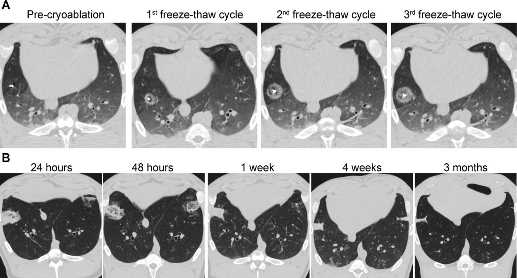
CT images showed that ablation zones with reduced transparency extended outward along the cryoprobe during the freeze-thaw cycles. Ground-glass opacity (GGO) attenuation was observed at the outer margin of the ablated lesions immediately after cryoablation (figure 2A). Figure 2B shows representative CT images of ablation zones during 3-month follow-up. The size of the ablated areas in four groups changed over time, which peaked at 24 hours and then gradually decreased (table 1, online supplemental table S1). Among all the groups, the ablation area of 15 min for three cycles was the largest, while the ablation area of 10 min for two cycles was the smallest. No significant difference was observed in the extent of the ablation zone between 10 min for three cycles and 15 min for two cycles (online supplemental figure S4).
Supplemental material

Representative CT findings of dynamic changes from the group 15 min for three cycles during cryoablation (A) and 3 months post cryoablation (B).
CT measurements of the ablated lesions (mean±SD)
Histopathology
24 hours post ablation
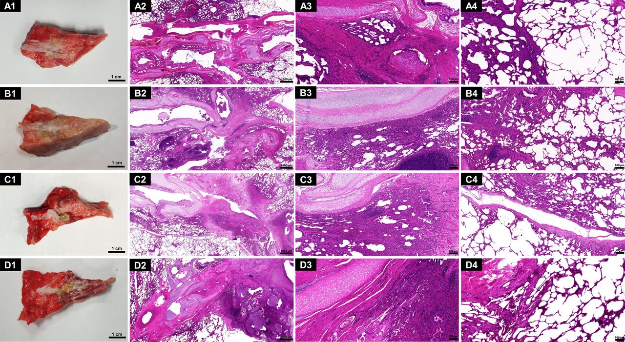
Macroscopically, the ablation zones were centred along the bronchus at the area of cryoprobe insertion, with a clear-cut edge that delineated dark red lesions inside the normal lung tissue (figure 3A1–D1), with an average volume of 5.11±1.18 cm3, 10.70±6.43 cm3, 8.98±7.71 cm3 and 14.08±9.20 cm3 (table 2, online supplemental table S2). H&E staining of the lung sections showed that the degree of injury was the most severe in the centre and gradually attenuated towards the periphery. The centre of the area of ablation perpendicular to the major axis of the target bronchus showed destruction of the bronchial epithelium, with leakage of red blood cells and fluid exudation. Importantly, cartilage structure remained intact (figure 3A2–D2). In subchondral lung tissues, the alveolar structure was destroyed, and the alveolar cavity was filled with massive red blood cells and fluid (figure 3A3–D3). Widened alveolar septa with congested capillaries and increased infiltration of inflammatory cells were noted in the area close to the peripheral lung tissue (figure 3A4–D4).

Representative histopathological findings at 24 hours after ablation in the groups 10 min for two cycles (A), 10 min for three cycles (B), 15 min for two cycles (C) and 15 min for three cycles (D). Gross pathology of the ablation zone along the bronchus at the area of cryoprobe insertion (A1–D1). H&E staining sections of the central ablation area perpendicular to the major axis of the target bronchus (A2–D2). H&E staining sections of the central ablation area around the cartilage (A3–D3) and the junction between the ablation zone and surrounding lung tissue (A4–D4). Scale bars: 1 cm for A1–D1; 1 mm for A2–D2, at 1.5× magnification; 100 µm for A3-4–D3-4, at 10× magnification.
Gross measurements of the ablated lesions (mean±SD)
4 weeks post ablation
Macroscopically, a grey-brown lesion was formed 4 weeks after the cryoablation (figure 4A1–D1). The size of the ablation zone significantly shrank compared with that in the 24-hour group. The average volumes were 0.21±0.23 cm3, 0.61±0.37 cm3, 1.02±0.78 cm3 and 1.37±1.11 cm3, respectively (table 2, online supplemental table S2). H&E staining showed markedly decreased acute haemorrhage and massive inflammation, accompanied by fibrous hyperplasia (figure 4A2–D2). The cartilage structure remained intact, but the arrangement seemed to be slightly disturbed and distorted (figure 4A3–D3). There was a gradual increase in inflammatory cell infiltration in the lung parenchyma, which was more severe than that observed in the 24-hour group (figure 4A4–D4). Masson staining showed the formation of fibrous tissues (online supplemental figure S5A,B).

Representative histopathological findings at 4 weeks after ablation in the groups 10 min for two cycles (A), 10 min for three cycles (B), 15 min for two cycles (C) and 15 min for three cycles (D). Gross pathology of the ablation zone along the bronchus at the area of cryoprobe insertion (A1–D1). H&E staining sections of the central ablation area perpendicular to the major axis of the target bronchus (A2–D2). H&E staining sections of the central ablation area around the cartilage (A3–D3) and the junction between the ablation zone and surrounding lung tissue (A4–D4). Scale bars: 1 cm for A1–D1; 1 mm for A2–D2, at 1.5× magnification; 100 µm for A3-4–D3-4, at 10× magnification.
3 months post ablation
Macroscopically, a long strip of greyish-yellow fibrous scar was formed 3 months after the cryoablation (figure 5A1–D1), in which the average volumes were 0.18±0.23 cm3, 0.69±0.37 cm3, 0.09±0.01 cm3 and 0.89±0.89 cm3, respectively (table 2, online supplemental table S2). H&E staining showed marked fibrous hyperplasia with chronic inflammation (figure 5A2–D2). The cartilage was structurally intact but disorganised and irregularly shaped (figure 5A3–D3) and surrounded by massive fibrous tissue (online supplemental figure S5C,D). Inflammatory cell infiltration consisted mainly of lymphocytes with lymphoid follicle formation (figure 5A4–D4).

Representative histopathological findings at 3 months after ablation in the groups 10 min for two cycles (A), 10 min for three cycles (B), 15 min for two cycles (C) and 15 min for three cycles (D). Gross pathology of the ablation zone along the bronchus at the area of cryoprobe insertion (A1–D1). H&E staining sections of the central ablation area perpendicular to the major axis of the target bronchus (A2–D2). H&E staining sections of the central ablation area around the cartilage (A3–D3) and the junction between the ablation zone and surrounding lung tissue (A4–D4). Scale bars: 1 cm for A1–D1; 1 mm for A2–D2, at 1.5× magnification; 100 µm for A3-4–D3-4, at 10× magnification.
Initial experience of transbronchial cryoablation in clinical practice
Patient and tumour characteristics
From 21 September 2022 to 3 February 2023, a total of nine patients with a mean age of 70±8 years have undergone transbronchial cryoablation. The baseline characteristics of the patients and tumours are summarised in table 3. All tumours had a definite pathological diagnosis prior to ablation. Nine adenocarcinomas, including one pulmonary metastasis from colon cancer, were treated with transbronchial cryoablation. Two tumours were located in the right upper lobes, three in the right lower lobe, two in the left upper lobes and two in the left lower lobe.
Patients and tumour characteristics receiving transbronchial cryoablation
Technical success and safety
A total of 10 cryoablations were successfully performed on 9 tumours, with the first case treated twice. The technical success rate is 100% (10/10). No procedure-related deaths occurred. One treatment-related complication occurred and was categorised as mild by the SIR classification system for complications.18 Mild thrombocytopaenia occurred in case 6 3 days after the cryoablation and recovered without any treatment within 1 month. No pneumothorax, haemorrhage or other postprocedural complications were observed. There were no changes in ECOG scores before and after the cryoablation. Moreover, the novel cryoprobe exhibited good operability and safety during the cryoablation, with no device-related adverse events reported. The working temperature was stable as illustrated in online supplemental figure S6. The temperature of the cryotip reached −150°C within 1.79±0.33 min, with a cooling rate of 85.06±14.14°C/min.
Efficacy
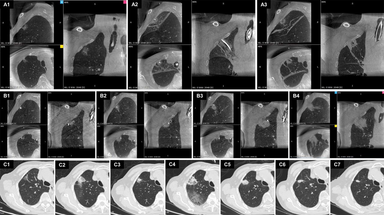
The procedures and representative imaging data of the first case of transbronchial cryoablation are shown in figure 6A,B. The patient underwent two cryoablations at a 1-month interval because of the tumour size and her advanced age. The target lesion was successfully covered in the ablation zone, and there was no evidence of haemorrhage or pneumothorax on CT. 1-day, 1-month, 3-month and the latest follow-up CT images of nine patients are shown in figures 6C and 7. Of the nine tumours, seven achieved complete ablation, while two exhibited incomplete ablation and subsequent local progression at 6 months (cases 3 and 4, figure 7B,C). The data from these nine cases yielded a technical efficacy of 77.78% and a disease control rate of 100% at 3 months.

Cone-beam CT (CBCT)-guided transbronchial cryoablation in case 1. CBCT images during the first cryoablation showing the target lesion before the procedure (A1), interprocedural confirmation of the cryoprobe inside the target lesion (A2) and the lesion immediately after cryoablation (A3). CBCT images of the second cryoablation showing the cryoprobe in three different target bronchi during cryoablation (B1–B3) and the lesion covered completely within the ablation area immediately after cryoablation (B4). Chest CT before cryoablation (C1), 1 day (C2) and 1 month (C3)after the first cryoablation and 1 day (C4), 1 month (C5), 3 months (C6) and 12 months (C7) after the second cryoablation. MPR, multiplanar reconstruction ; WL, window level; WW, window width.

Chest CT before cryoablation (A1–H1), 1 day (A2–H2), 1 month (A3–H3), 3 months (A4–H4) and 6–12 months (A5, D5 and E5, 12-month follow-up; F5, G5 and H5, 9-month follow-up; and B5 and C5, 6-month follow-up) after the transbronchial cryoablation in cases 2–9.
Discussion
Image-guided ablation is recommended as a non-surgical treatment for patients who are not surgical candidates because of medical comorbidities. The advantages of local tumour ablation over surgery and radiotherapy include lung preservation and the ability to repeat treatments.19 A retrospective study reported that RFA had similar overall survival and progression-free survival to surgery in the treatment of solitary or multiple lung metastases measuring <4 cm in diameter.20 Unlike heat-based ablation, cryoablation can be safely performed in high oxygen settings,9 as well as for tumours near the great vessels, pericardium, pleura or fissures.21–23 As shown in cases 1 and 5, transbronchial cryoablation was successfully performed on tumours adjacent to the pleura and interlobar fissure without any severe pain or other complications during and after the procedure. Moreover, the cryoablation zone can be monitored by direct CT monitoring of ice ball formation,21 the border of which represents a temperature of 0℃. This can help to determine the underlying cytotoxic isotherm (ie, −20°C isotherm), locating approximately 5 mm within the 0°C isotherm,24–26 which can be clinically useful during intraprocedural monitoring of the zone of ablation on CT or CBCT. Several studies have shown that the −20°C isotherm was most closely related to the pathological zone of complete necrosis.25 26 Hence, we should ensure that the edge of the ice ball (0°C) observed on CT or CBCT is at least 5 mm beyond that of the tumour so as to avoid undertreatment in clinical practice.
An additional promising advantage of cryoablation is that it may favour a specific immune response against tumours, which may stimulate or enhance the combined efficacy of antitumour immunotherapy.27–31 Compared with the percutaneous approach, the transbronchial approach seems to be a promising therapeutic alternative with a lower risk of pneumothorax.9 32 Previous studies have reported that transbronchial RFA and MWA are feasible for the treatment of early-stage peripheral lung cancer.12–14 33 However, to our knowledge, bronchoscopy-guided cryoablation in clinical practice has not been investigated to date.
The entire animal experiment was performed under CT monitoring, and evolution of the ablation zone was observed at 24 hours, 48 hours, 1 week, 4 weeks and 3 months after cryoablation. The change in the ablation zones on CT was similar to that in our previous study.16 The gross anatomy and pathology at 24 hours, 4 weeks and 3 months after cryoablation showed a change from massive haemorrhage to fibrous hyperplasia accompanied by chronic inflammation, which indicated the repair process after cryoablation consistent with CT images. Most importantly, the bronchial cartilage remained intact. In addition to extending the follow-up time to 3 months, we also compared the differences between four experimental groups with different freezing time (10 min and 15 min) and the number of cycles (two and three). The extent of ablation increased with freezing time and the number of freeze-thaw cycles, which is consistent with previous studies.34 35
Unlike animal experiments in which a predetermined cryoablation probe can be placed in the centre of the lesion to completely cover the target lesion, the cryoprobe may fail to reach the centre of the lesion in clinical practice. This is because the target bronchus does not lead to the centre of the lesion, or the lesion is too large. Thus, conformal ablation in multiple points of one bronchus or multiple bronchi leading to or adjacent to the target lesion is needed, aiming to use multiple overlapping treatments to completely cover the target lesion. To achieve this goal, comprehensive preoperative planning and continuous monitoring during ablation are required. The crucial issue is the precision of the introduction of the ablation catheter into the tumour. Navigation bronchoscopy can help to offer the ability to accurately reach the target tumour. Before ablation, the spatial relationship between the bronchus and the lesion should be confirmed at different three-dimensional levels to determine the target bronchus to be ablated. Another concern for transbronchial ablation is simultaneous visualisation of probe delivery and intraprocedural assessment of the ablation efficacy.14 From our experience, the use of CBCT imaging to assess probe positioning with respect to the targeted lesion36 and size of the ablation zone37 was beneficial and should be considered an integral part of transbronchial cryoablation. In addition, good ventilation is required to prevent the occurrence of pulmonary atelectasis because of the long operation time of cryoablation. Otherwise, it will affect the judgement of the position of the probe and the lesion, as well as monitoring ablation efficacy.
No procedure-related deaths occurred in our clinical study, and only one mild treatment-related complication occurred. The first four patients were treated with a freeze-thaw time of 7 min and 3 min, respectively, for each cycle of the ablation due to safety concerns. Among them, cases 3 and 4, both with solid nodules, exhibited local progression in the 6-month follow-up CT scans. This prompted us to consider inadequate freezing time as a potential factor contributing to incomplete ablation. Moreover, the variability in pulmonary nodules among different patients demands more individualised treatments in clinical settings. Case 3, for instance, was diagnosed as pulmonary metastasis from colon cancer, indicating a higher degree of malignancy and possibly requiring longer freezing time or additional freeze-thaw cycles. Continued experience is still needed to develop appropriate procedures for safe and effective treatment in clinical practice.
Nowadays, various guided bronchoscopy techniques have extended the range of pulmonary lesions that can be accessed by bronchoscopy. Bronchoscopic transparenchymal nodule access can allow access to a pulmonary lesion without an endobronchial route by creating a tunnel through the lung parenchyma directly into the lesion.38 Besides, robotic bronchoscopy has emerged as a novel localisation technique of peripheral pulmonary lesions with greater stability and accuracy. In the future, these techniques should be combined with transbronchial ablation to benefit more patients.
This study still had several limitations. First, transbronchial cryoablation was performed in normal porcine lungs without tumours. However, the size of the ablation zones in animal experiments may serve as a lower limit for the expected extent of ablation because normal lung tissues contain more air than lung tumours and thus have worse heat conductivity.39 40 Furthermore, normal lung tissues may resemble the GGO component in a lung nodule on CT because of the preservation of the alveolar structure of the cancer cells in GGO.41 In the future, standardised large animal models of lung tumours should be established to provide more relevant data. Second, the animal experiment showed that the size of cryoablation zone was smaller than that in the previous experiment,16 because the novel cryoprobe used in this study was thinner than the previous one so that it could reach more peripheral lesions. To solve this problem, we applied multiple overlapping cryoablation in clinical practice to obtain larger tumour coverage. Last but not least, the number of patients enrolled was small, and the follow-up was short so far. Hence, long-term follow-up and a larger cohort are still needed to determine the efficacy of transbronchial ablation for early-stage NSCLC.
Conclusion
Our initial experience suggested that transbronchial cryoablation with a novel thin cryoprobe is a feasible and safe treatment for early-stage NSCLC patients or metastases. Further studies with larger cohorts and longer follow-up are warranted to confirm its clinical efficacy.
Supplemental material
Data availability statement
Data are available upon reasonable request.
Ethics statements
Patient consent for publication
Ethics approval
This study involves human participants. The prospective single-arm clinical trial was approved by the Ethics Committee at Shanghai Chest Hospital (Shanghai, China) (LS22027). Participants gave informed consent to participate in the study before taking part. The protocol for the animal study was approved by the Ethics Committee at Shanghai Chest Hospital (Shanghai, China) (KS21020) and Gateway Medical Innovation Center (Shanghai, China) (SH2021-09001).
Acknowledgments
The authors thank Xia Tong for her help in the clinical trial registry application.
References
Supplementary materials
Supplementary Data
This web only file has been produced by the BMJ Publishing Group from an electronic file supplied by the author(s) and has not been edited for content.
Footnotes
CG, HY and CY contributed equally.
Contributors JS is the guarantor of the content of the manuscript and takes responsibility for the content of the manuscript, integrity of the data and the accuracy of the data analysis. CG and HY contributed to the experimental design, experimental operation, data collection, data interpretation, pathological analysis, writing of the manuscript and manuscript review. CY contributed to the experimental design, experimental operation, data collection and manuscript review. FX and JC contributed to the data interpretation and manuscript review. LZ contributed to the pathological analysis and manuscript review. YJ contributed to the data interpretation and manuscript review.
Funding This study was supported by the National Multi-disciplinary Treatment Project for Major Diseases (2020NMDTP) and Clinical Research Plan of SHDC (SHDC2020CR3081B).
Competing interests Dr Chi Yang is a full-time employee of AccuTarget MediPharma (Shanghai) Co., Ltd. Dr Jiayuan Sun serves on the advisory abroad and receives consultant fees from AccuTarget MediPharma (Shanghai) Co., Ltd. The other authors declare no competing interests.
Provenance and peer review Not commissioned; externally peer reviewed.
Supplemental material This content has been supplied by the author(s). It has not been vetted by BMJ Publishing Group Limited (BMJ) and may not have been peer-reviewed. Any opinions or recommendations discussed are solely those of the author(s) and are not endorsed by BMJ. BMJ disclaims all liability and responsibility arising from any reliance placed on the content. Where the content includes any translated material, BMJ does not warrant the accuracy and reliability of the translations (including but not limited to local regulations, clinical guidelines, terminology, drug names and drug dosages), and is not responsible for any error and/or omissions arising from translation and adaptation or otherwise.